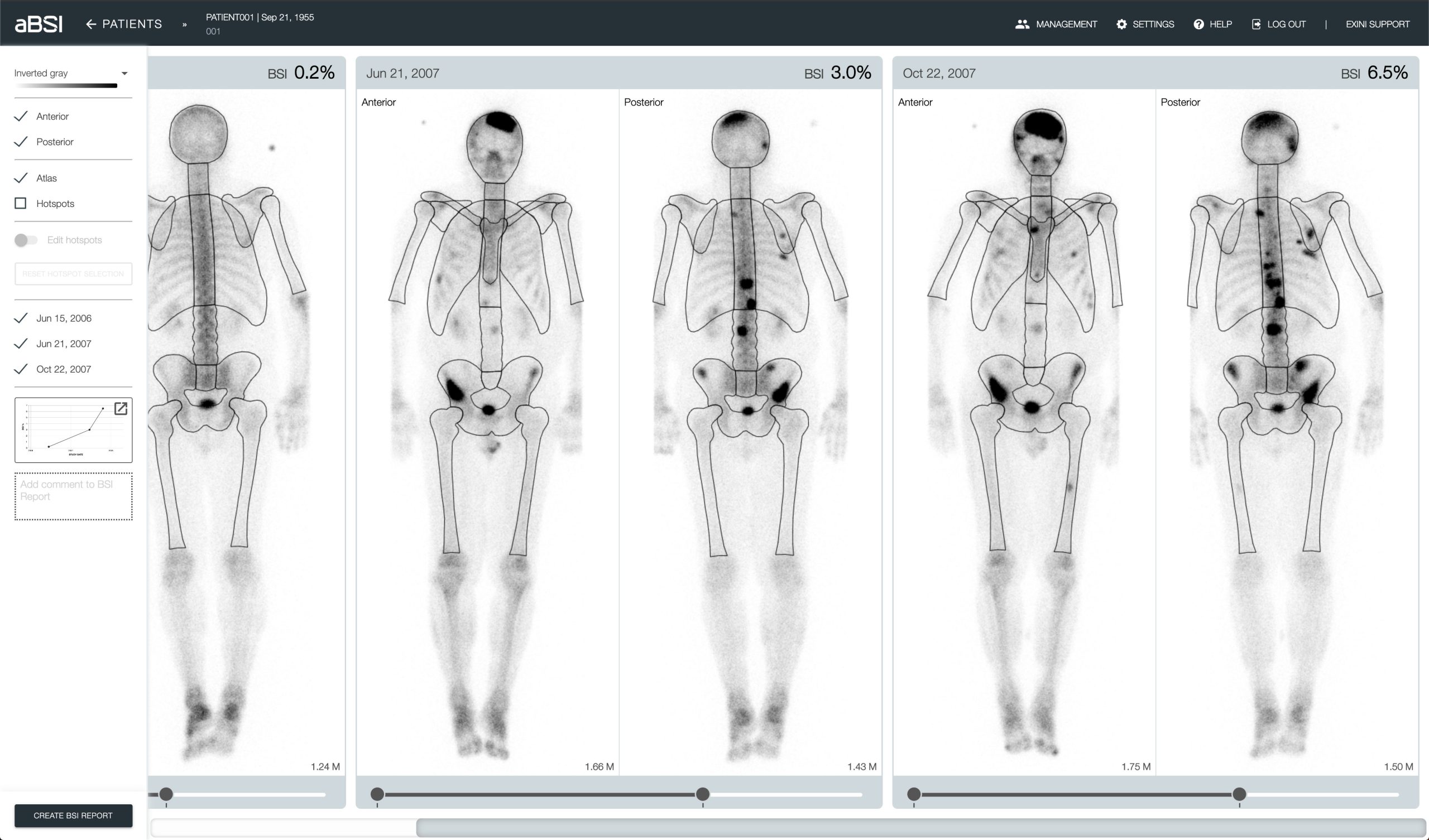
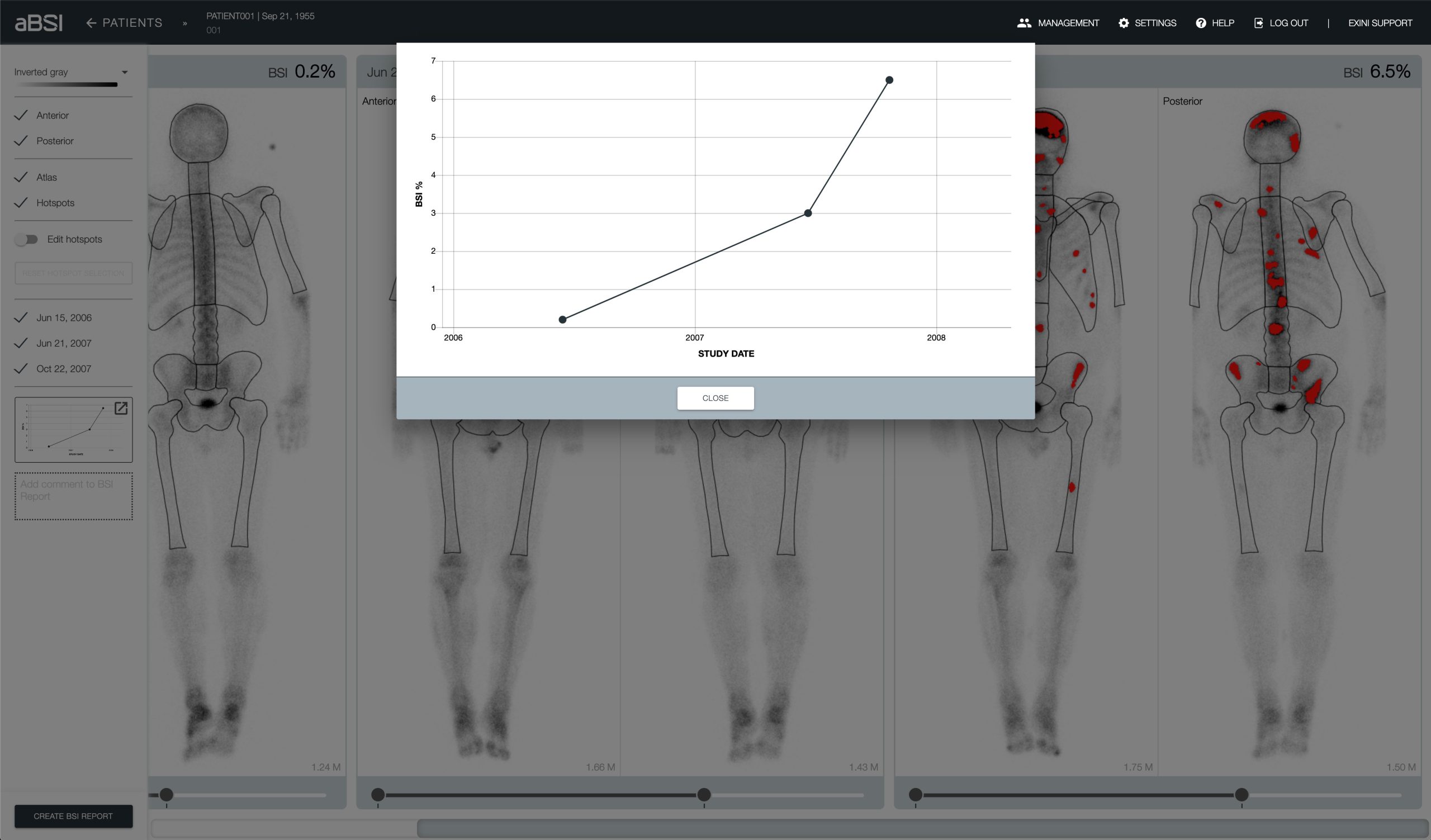
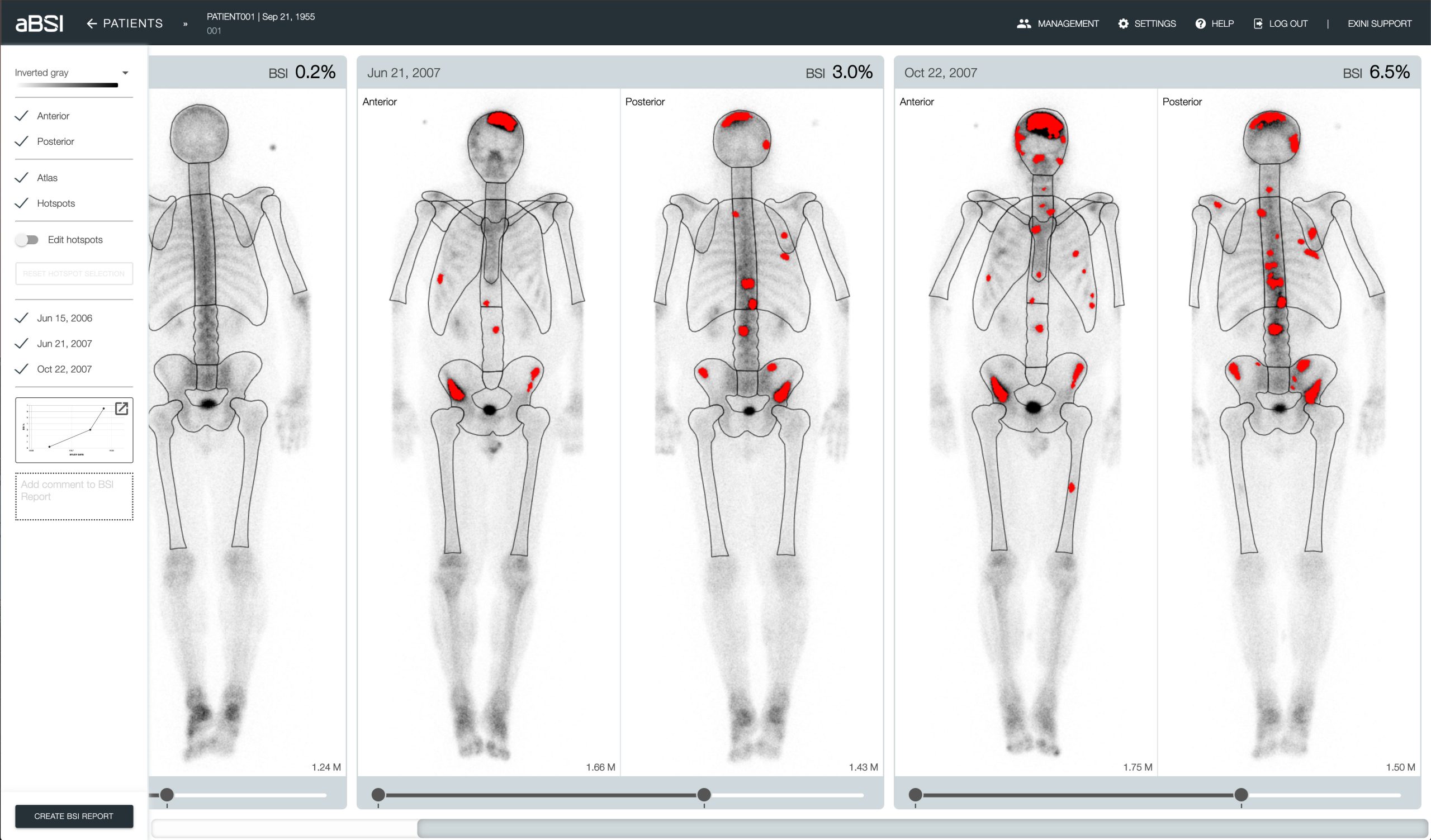
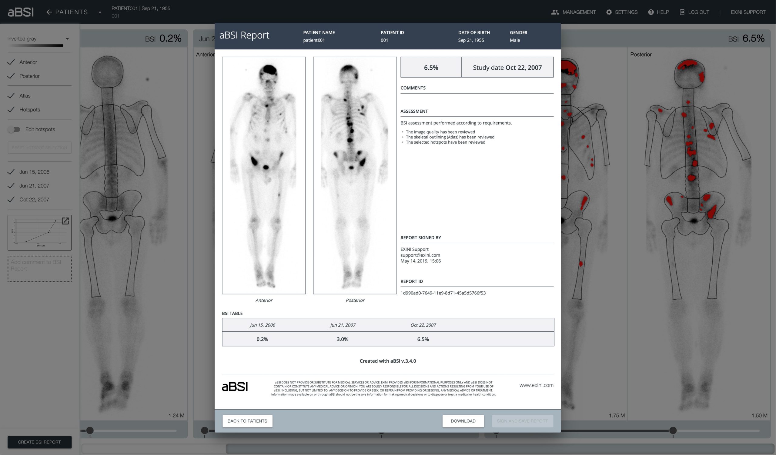
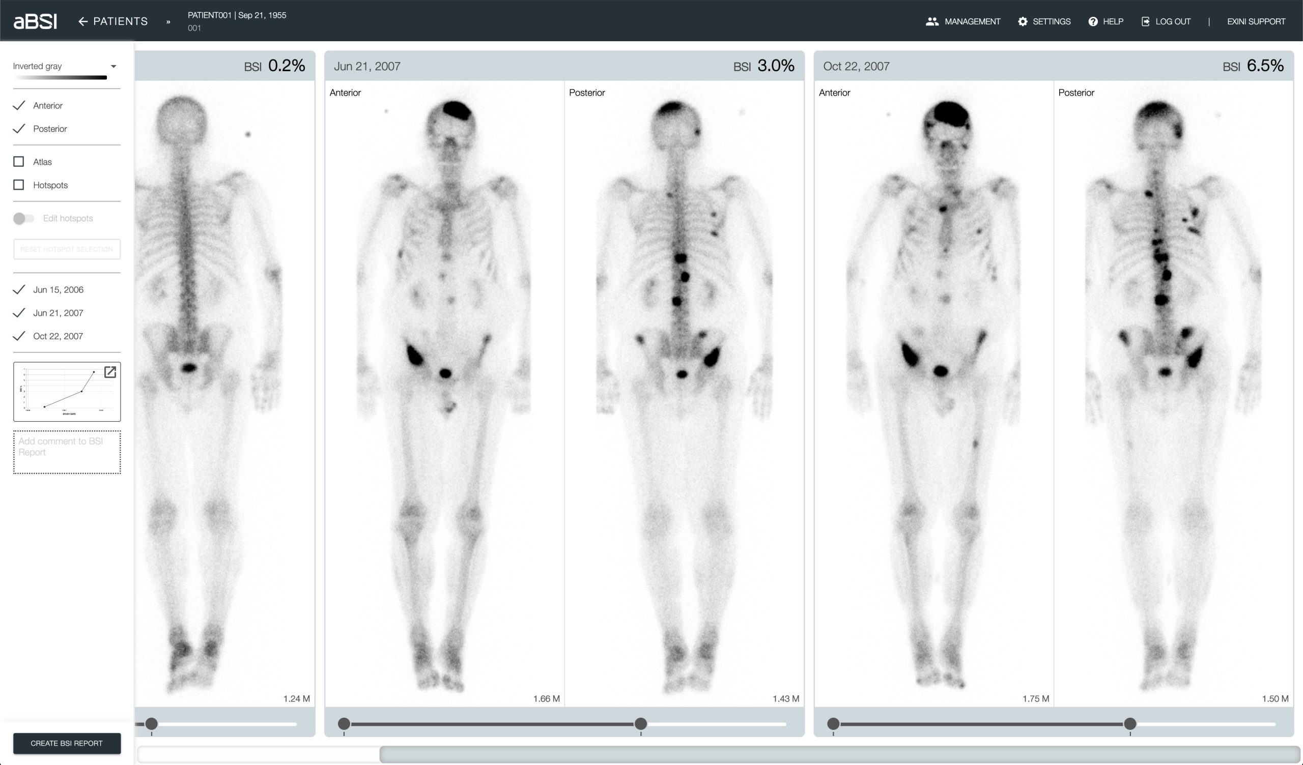
The automated Bone Scan Index (aBSI) technology, developed by EXINI Diagnostics AB, gives a fully quantitative assessment of bone scans that incorporates inferred masses of all lesions. The computed index reflects the proportion of the total skeleton mass that is observed to have tumor involvement1. In a prospectively defined multi-institutional phase 3 study with 721 metastatic prostate cancer patients, aBSI was found to be an independent prognostic determinant of overall survival and the study supported using aBSI in the design and eligibility for clinical trials for systemic therapies for metastatic castration-resistant prostate cancer2.

As of 2017, aBSI is provided as a secure and compliant web application readily available from a web browser by logging in to www.bonescanindex.com.  In Europe, the device is available for clinical use in Austria, Denmark, France, Germany, Ireland, Netherlands, Sweden, Switzerland and United Kingdom. As of October 2019, aBSI is also available for clinical use in the United States with a 510(k) clearance (K191262).

aBSI is also available for local installation under the name EXINI bone. As of April 2022, EXINI bone is CE marked according to MDR 2017/745

  1. Ulmert D, et al. A novel automated platform for quantifying the extent of skeletal tumour involvement in prostate cancer patients using the Bone Scan Index. Eur Urol. 2012.
  2. Armstrong AJ et al. Phase 3 Assessment of the Automated Bone Scan Index as a Prognostic Imaging Biomarker of Overall Survival in Men With Metastatic Castration-Resistant Prostate Cancer: A Secondary Analysis of a Randomized Clinical Trial. JAMA Oncol. 2018.

GDPR

When you use aBSI to process patient information related to a patient who is a resident of the EU, you are responsible for ensuring that your organization complies with GDPR. In terms of GDPR you, as the user of aBSI, are the data controller and EXINI, as the service provider, is the data processor. In advance of processing data with aBSI, be sure that you have explicit consent from the patient whose data you are capturing. When data is sent to aBSI, it is stored in a secure manner, and is encrypted in transit and at rest.

Our Commitment

EXINI (‘we’ or ‘us’ or ‘our’) are committed and dedicated to ensuring the security and protection of the personal information that we process, and to provide a robust, continuous and consistent approach to data protection. Our objectives for GDPR and HIPAA compliance include the development and implementation of data protection roles, policies, procedures, controls and measures to ensure continuous safeguarding of the personal information under our remit.

How we are implementing GDPR and HIPAA

  • Policies & Procedures – Data protection policies and procedures to meet the requirements and standards of the GDPR and any relevant data protection laws, including HIPAA, are in place
  • Data Retention & Erasure – we have retention policies and are applying the privacy by design principle, meaning we store only data that is needed for the current task and only store it for as long as needed
  • Data Breaches – as a medical device manufacturer we have breach procedures in place that ensure safeguards and measures to identify, assess, investigate and report any personal data breach at the earliest possible time
  • International Data Transfers & Third-Party Disclosures – where EXINI stores or transfers personal information outside the EU, we have robust procedures and safeguarding measures in place to secure, encrypt and maintain the integrity of the data
  • Processor Agreements – where we use a third-party to process personal information on your behalf we have data processor agreements and/or business associate agreements in place

BONENAVI

BONENAVI, the platform to calculate bone scan index in Japan, was launched 2011 with partner FUJIFILM RI Pharma. The platform is developed and validated for Japanese patient population. Since its launch in 2011, nearly 900 hospitals have incorporated BONENAVI in their regular clinical workflow. Illustrative BONENAVI reports are used in discussing treatment evaluation with patients. The society of nuclear medicine in Japan recommends bone scan index for use in metastatic prostate cancer to assess metastatic burden in bone. The availability of BONENAVI has generated clinical value of the bone scan in prostate cancer.

In June 2019, FUJIFILM bought the exclusive rights of BONENAVI in Japan. 

Explore aBSI

Use aBSI

Log in

If you already have an account, you can use it to log in to aBSI from your computer.

Publications

Our technology, included in our product aBSI and BONENAVI, is well documented in several studies.

Help

Here you can find the most frequently asked questions and contact information to our customer support.

User manuals – EXINI bone

aBSI and EXINI bone

A software as a medical device manufactured by EXINI Diagnostics AB.

Performance Characteristics

In non-clinical and clinical investigations, aBSI/EXINI bone has demonstrated safety and effectiveness in analyzing and quantifying nuclear medicine bone scan in oncology. In a set of analytical validation studies demonstrated the following: Linearity of the automated BSI interpretations in the range of 0.10–13.0 was confirmed, and Pearson correlation was observed at 0.995 (n=50; 95% confidence interval, 0.99–0.99; P<0.0001). The mean coefficient of variation was less than 20%. The mean BSI difference between the 2 repeat bone scans of 35 patients was 0.05 (SD=0.15), with an upper confidence limit of 0.30. The interobserver agreement in the automated BSI interpretations was more consistent (κ=0.96, P<0.0001) than the qualitative visual assessment of the changes (κ=0.70, P<0.0001) was in the bone scans of 173 patients. In a large prospectively phase III clinical study, investigators demonstrated that in a multivariable survival model, aBSI/EXINI bone remained independently associated with OS (HR, 1.06; 95% CI, 1.01- 1.11; P=0.03).

Warnings

Always ensure that the patient name, patient ID and study date displayed in the patient section correspondto the patient case you intend to review.

The quantification results including the automated BSI must not be used as a tool for diagnostic or treatment decisions. They are intended to be used as a guide in the visual interpretation of bone scans. You must not rely solely on the quantification results of the software but use additional data points to determine a diagnosis and necessary treatment.

Always review the images and quantification results in the BSI report to ensure that the information exported is correct.

Ydelsesspecifikationer

I ikke-kliniske og kliniske undersøgelser har aBSI/EXINI bone vist sig at kunne analysere og kvantificere nuklearmedicinsk knoglescanning inden for onkologi på en sikkert og effektiv måde. En række analytiske valideringsundersøgelser viste følgende: Lineariteten af de automatiserede BSI-fortolkninger i intervallet 0,10-13,0 blev bekræftet, og Pearson-korrelationen blev observeret ved 0,995 (n=50; 95% konfidensinterval, 0,99-0,99; P<0,0001). Den gennemsnitlige variationskoefficient var mindre end 20 %. Den gennemsnitlige BSI-forskel mellem to gentagne knoglescanninger af 35 patienter var 0,05 (SD=0,15) med en øvre konfidensgrænse på 0,30. Interobserveroverensstemmelsen i de automatiserede BSI-fortolkninger var mere konsistente (κ= 0,96, P<0,0001), end den kvalitative visuelle vurdering af ændringerne (κ= 0,70, P=0,0001) var i knoglescanninger af 173 patienter. I en stor prospektiv fase III klinisk undersøgels påviste forskerne, at i en multivariabel overlevelsesmodel forblev aBSI/EXINI bone uafhængigt forbundet med OS (HR, 1,06; 95% CI, 1,01-1,11; P = 0,03).

Advarsler

Det skal altid sikres, at patientens navn, patient-ID og studiets dato, der vises i patientafsnittet, svarer til den patient, der skal gennemgås.

Kvantificeringsresultaterne, herunder den automatiske BSI-værdi, må ikke anvendes til diagnostiske eller behandlingsmæssige beslutninger. Disse resultater er beregnet til at blive brugt vejledende ved den visuelle fortolkning af knoglescanninger. Undlad udelukkende at stole på kvantificeringsresultaterne fra softwaren, men brug yderligere datapunkter til at finde frem til en diagnose og den tilhørende behandling.

Gennemgå altid billederne og kvantificeringsresultaterne i BSI-rapporten for at sikre, at de eksporterede oplysninger er korrekte.

Leistungsmerkmale

aBSI/EXINI bone hat in nicht-klinischen und klinischen Studien Sicherheit und Wirksamkeit bei der Analyse und Quantifizierung des nuklearmedizinischen Knochenscans in der Onkologie gezeigt. In einer Reihe von analytischen Validierungsstudien wurde Folgendes nachgewiesen: Die Linearität der automatisierten BSI-Interpretationen im Bereich von 0,10–13,0 wurde bestätigt, und die Pearson-Korrelation wurde bei 0,995 beobachtet (n=50; 95% Konfidenzintervall, 0,99–0,99; P<0,0001). Der mittlere Variationskoeffizient lag unter 20 %. Die mittlere BSI-Differenz zwischen den 2 wiederholten Knochenscans von 35 Patienten betrug 0,05 (SD=0,15) mit einer oberen Konfidenzgrenze von 0,30. Die Interobserver-Übereinstimmung in den automatisierten BSIInterpretationen war konsistenter (κ=0,96, P<0,0001) als die qualitative visuelle Beurteilung der Veränderungen (κ=0,70, P<0.0001) bei den Knochenscans von 173 Patienten. In einer großen prospektiven klinischen Phase-III-Studie zeigten die Forscher, dass aBSI/EXINI bone in einem multivariablen Überlebensmodell unabhängig mit OS assoziiert blieb (HR 1,06; 95% CI, 1,01-1,11; P=0,03).

Warnungen

Vergewissern Sie sich immer, dass der Patientenname, die Patienten-ID und das Studiendatum, die im Patientenbereich angezeigt werden, mit dem Patientenfall, den Sie prüfen möchten, übereinstimmen.

Die Quantifizierungsergebnisse einschließlich des automatisierten BSI dürfen nicht als Grundlage für Diagnose- oder Behandlungsentscheidungen herangezogen werden. Sie sollen als Richtlinie für die visuelle Interpretation von Knochenscans dienen. Stützen Sie sich nicht ausschließlich auf die Quantifizierungsergebnisse der Software, sondern verwenden Sie zusätzliche Datenpunkte, um eine Diagnose zu erstellen und die erforderliche Behandlung festzulegen.

Prüfen Sie immer die Bilder und Quantifizierungsergebnisse im BSI-Bericht, um sicherzustellen, dass die exportierten Informationen korrekt sind.

Características de rendimiento

En investigaciones no clínicas y clínicas, aBSI/EXINI bone ha demostrado seguridad y eficacia en el análisis y cuantificación de la gammagrafía ósea de medicina nuclear en oncología. En un conjunto de estudios de validación analítica demostró lo siguiente: Se confirmó la linealidad de las interpretaciones automatizadas del BSI en el rango de 0,10–13,0, y se observó la correlación de Pearson a 0,995 (n=50; 95 % intervalo de confianza, 0,99–0,99; P<0,0001). El coeficiente de variación medio fue inferior al 20 %. La diferencia media del BSI entre las dos gammagrafías óseas repetidas de 35 pacientes fue de 0,05 (SD=0,15), con un límite de confianza superior de 0,30. La concordancia entre observadores en las interpretaciones automatizadas del BSI fue más coherente (κ=0,96, P<0,0001) que la evaluación visual cualitativa de los cambios (κ= 0,70, P<0,0001) se encontraba en las gammagrafías óseas de 173 pacientes. En un estudio clínico grande prospectivo en fase III, los investigadores demostraron que en un modelo de supervivencia multivariable, aBSI/EXINI bone permaneció asociado independientemente con OS (HR, 1,06; IC del 95 %, 1,01-1,11; P=0.03).

Advertencias

Asegúrese siempre de que el nombre del paciente, el identificador del paciente y la fecha del estudio que se muestran en la sección del paciente se correspondan con el caso del paciente que pretende revisar.

Los resultados de cuantificación, incluido el BSI automatizado, no deben utilizarse como herramienta para decisiones de diagnóstico o tratamiento. Están destinados a ser utilizados como una guía en la interpretación visual de las gammagrafías óseas. No debe basarse únicamente en los resultados de cuantificación del software, sino utilizar puntos de datos adicionales para determinar un diagnóstico y el tratamiento necesario.

Revise siempre las imágenes y los resultados de cuantificación en el informe del BSI para asegurarse de que la información exportada sea correcta.

Caractéristiques de performance

Dans le cadre d’études non cliniques et cliniques, aBSI/EXINI bone a démontré sa sécurité et son efficacité dans l’analyse et la quantification de la scintigraphie osseuse (médecine nucléaire) en oncologie. Un ensemble d’études de validation analytique a démontré les points suivants : la linéarité des interprétations automatisées du BSI dans la plage de 0,10-13,0 a été confirmée, et la corrélation de Pearson a été observée à 0,995 (n=50 ; intervalle de confiance à 95 %, 0,99-0,99 ; P<0,0001). Le coefficient de variation moyen était inférieur à 20 %. La différence de BSI moyenne entre les 2 scintigraphies osseuses consécutives chez 35 patients était de 0,05 (ET=0,15), avec une limite de confiance supérieure de 0,30. La concordance inter-observateur dans les interprétations automatisées du BSI était plus cohérente (κ=0,96, P<0,0001) que l’évaluation visuelle qualitative des variations (κ=0,70, P< 0,0001) dans les scintigraphies osseuses de 173 patients. Dans une vaste étude clinique prospective de phase III, les investigateurs ont démontré, dans un modèle de survie multivarié, que le aBSI/EXINI bone automatisé est resté indépendamment associé à la SG (HR, 1,06 ; IC à 95 %, 1,01-1,11 ; P = 0,03).

Mises en garde

Vérifiez toujours que le nom du patient, son identifiant et la date de l’étude indiqués dans la section du patient correspondent bien au cas du patient que vous souhaitez examiner.

Les résultats de quantification, y compris le BSI automatisé, ne doivent pas être utilisés pour la prise de décisions diagnostiques ou thérapeutiques. Ils sont prévus pour servir de guide dans l’interprétation visuelle des scintigraphies osseuses. Vous ne devez pas vous appuyer uniquement sur les résultats de quantification du logiciel, mais utiliser également des points de données supplémentaires afin de poser un diagnostic et de déterminer le traitement approprié.

Passez toujours en revue les images et les résultats de quantification dans le rapport de BSI pour vous assurer que les informations exportées sont correctes.

Prestatiekenmerken

In niet-klinische en klinische onderzoeken heeft aBSI/EXINI bone de veiligheid en effectiviteit aangetoond bij het analyseren en kwantificeren van botscans door nucleaire geneeskunde in de oncologie. In een reeks analytische valideringsstudies is het volgende aangetoond: De lineariteit van de geautomatiseerde BSI-interpretaties in het bereik van 0,10-13,0 werd bevestigd en Pearsoncorrelatie werd waargenomen op 0,995 (n=50; 95% betrouwbaarheidsinterval, 0,99-0,99; P<0,0001). De gemiddelde variatiecoëfficiënt was minder dan 20%. Het gemiddelde BSI-verschil tussen de 2 herhaalde botscans van 35 patiënten was 0,05 (SD=0,15), met een bovengrens van de betrouwbaarheid van 0,30. De interobserverovereenkomst in de geautomatiseerde BSI-interpretaties was consistenter (κ= 0,96, P<0,0001) dan de kwalitatieve visuele beoordeling van de veranderingen (κ=0,70, P<0,0001) in de botscans van 173 patiënten. In een grote prospectieve fase III klinische studie hebben onderzoekers aangetoond dat in een multivariabel survivalmodel, aBSI/EXINI bone onafhankelijk geassocieerd bleef met OS (HR, 1,06; 95% BI, 1,01-1,11; P = 0,03).

Waarschuwingen

Controleer altijd of de naam van de patiënt, patiënt-ID en onderzoeksdatum die in de patiëntensectie worden weergegeven, overeenkomen met het patiëntendossier dat u gaat bekijken.

De gekwantificeerde resultaten inclusief de automatische BSI mogen niet worden gebruikt als hulpmiddel voor diagnostiek of bij behandelbeslissingen. Ze zijn bedoeld als handleiding voor de visuele interpretatie van botscans. U mag niet uitsluitend vertrouwen op de gekwantificeerde resultaten van de software, maar u dient extra gegevenspunten te gebruiken om een diagnose te stellen en een noodzakelijke behandeling te bepalen.

Bestudeer altijd de beelden en kwantificeringsresultaten in het BSI-rapport om te zorgen dat de informatie die wordt geëxporteerd correct is.

Ytelsesegenskaper

I ikke-kliniske og kliniske undersøkelser har aBSI/EXINI bone vist seg å være sikker og effektiv ved analysering og kvantifisering av nukleærmedisinsk skjelettscintigrafi for onkologi. Analytiske valideringsstudier viste følgende: Lineariteten til de automatiserte BSItolkningene i området 0,10–13,0 ble bekreftet, og Pearson korrelasjonskoeffesient var 0,995 (n=50; 95 % konfidensintervall, 0,99–0,99; P<0,0001). Gjennomsnittlig variasjonskoeffisient var mindre enn 20 %. Gjennomsnittlig BSI-forskjell mellom de to gjentatte skjelettscintigrafiene av 35 pasienter var 0,05 (SD=0,15), med en øvre konfidensgrense på 0,30. Overensstemmelse mellom observatører for de automatiserte BSI-tolkningene var bedre (κ=0,96, P<0,0001) enn for den kvalitative visuelle vurderingen av endringene (κ=0,70, P<0,0001) i skjelettscintigrafier av 173 pasienter. I en stor klinisk prospektiv fase III-studie viste utprøvere at aBSI/EXINI bone forble uavhengig assosiert med OS i en multivariabel overlevelsesmodell (HR, 1,06; 95 % KI, 1,01–1,11; P=0,03).

Advarsler

Sørg alltid for at pasientnavn, pasient-ID og studiedatoen som vises i pasientdelen, stemmer med det pasienttilfellet du skal granske.

Kvantifiseringsresultatene, som omfatter den automatiserte BSI (Bone Scan Index), må ikke brukes som et verktøy for beslutninger om diagnose eller behandling. De er ment å brukes som veiledning ved visuell tolkning av skjelettscintigrafier. Du må ikke stole utelukkende på programvarens kvantifiseringsresultat, men benytte ytterligere datapunkter for å faststille en diagnose og nødvendig behandling.

Du må alltid granske bildene og kvantifiseringsresultatene i BSI-rapporten for å sikre at den eksporterte informasjonen er korrekt.

Prestandaegenskaper

I icke-kliniska och kliniska undersökningar har aBSI/EXINI bone visat säkerhet och effektivitet i att analysera och kvantifiera nukleärmedicinska skelettscintigrafier inom onkologi. I en uppsättning analytiska valideringsstudier visades följande:
Linjäriteten hos de automatiserade BSI-tolkningarna i intervallet 0,10–13,0 bekräftades, och Pearson-korrelationen observerades vid 0,995 (n=50; 95 % konfidensintervall, 0,99–0,99; P<0,0001). Den genomsnittliga variationskoefficienten var mindre än 20 %. Den genomsnittliga BSI-skillnaden mellan 2 upprepade skelettscintigrafier av 35 patienter var 0,05 (SD=0,15), med ett övre konfidensintervall på 0,30. Interobserveravtalet i de automatiserade BSI-tolkningarna var mer konsekventa (κ=0,96, P<0,0001) än vad den kvalitativa visuella bedömningen av förändringarna (κ=0,70, P<0,0001) var i skelettscintigrafierna av 173 patienter. I en stor prospektiv klinisk fas III-studie, har utredarna visat att aBSI/EXINI bone, i en multivariabel överlevnadsmodell, förblev oberoende relaterad till OS (HR, 1,06; 95 % CI, 1,01-1,11; P = 0,03).

Varningar

Säkerställ alltid att patientnamn, patient-ID och studiedatum som visas i patientdelen verkligen stämmer med det patientfall du ska granska.

Kvantifieringsresultaten, som innefattar den automatiserade BSI (Bone Scan Index), får inte användas som ett verktyg för beslut om diagnos eller behandling. De är avsedda att användas som vägledning vid visuell tolkning av skelettscintigrafier. Du får inte lita enbart till programvarans kvantifieringsresultat, utan måste använda ytterligare datapunkter för att fastställa en diagnos och nödvändig behandling.

Granska alltid bilderna och kvantifieringsresultaten i BSI-rapporten för att säkerställa att den exporterade informationen är korrekt.

Trademark

EXINI® is a Registered Trademark.

Disclaimer

The materials on this website are of a general, informational nature. The content does not purport to be exhaustive and should not be relied upon as a substitute or replacement for individual medical, technical or instructional advice on any specific matter. If you have a specific question, you are most welcome to address it to one of our representatives or through the appropriate e-mail. Links to other websites are for information only. We do not endorse or accept any responsibility or liability for the material on any website referred to or accessed through this website. All links to our website should go to the top-level page.